Management of Peri-implantitis on a Smooth-surfaced Implant
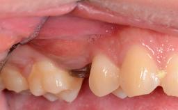
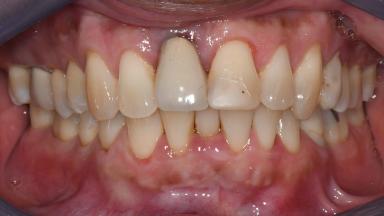
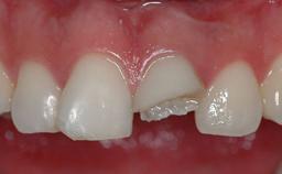
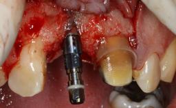
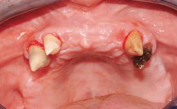
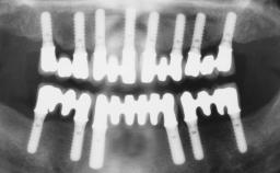
With this case, Xian Jun Edwin Goh demonstrates how proper diagnosis of an implant related complication is critical in successful management of peri-implantitis. A 56 years old male patient presented with peri-implantitis related to the dental implant replacing his upper right central incisor (11 site). Following proper diagnosis and non-surgical periodontal treatment, surgical intervention consisting of open flap debridement and connective tissue graft was performed. The patient was then enrolled in an individualised maintenance regime and continued to demonstrate stable periodontal and peri-implant parameters.

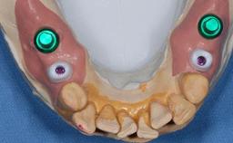
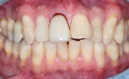
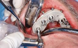
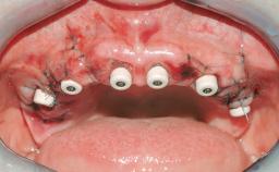
General Risk Assessment
Patient-related Factors
| Smoking Habit | None |
|---|---|
| Oral hygiene | Poor |
| Compliance | Adequate |
| Patient's Expectations | Realistic |
Patient-medical Factors
| Medical Status | Healthy, uneventful healing |
|---|---|
| Medical Fitness | Moderately compromised health, but can undergo planned anesthesia and surgical procedure (ASA II to ASA III) |
| Medications | No medications that would negatively affect the surgical procedure and outcomes. |
| Radiation Treatment | None |
| Growth Status | Complete |
Site-related Factors
| Periodontal Status | Active periodontal disease. |
|---|---|
| Access | Adequate |
| Pathology near the implant site | None |
| Previous surgeries in planned implant site | Previous procedures resulting in none or minimal bone and soft tissue changes. |
Surgical Classification
Surgical Complexity
| Timing of placement | Healed (Type IV) |
|---|---|
| Simultaneous or Staged grafting procedures | Implant placement with simultaneous hard and soft tissue procedures |
Anatomy
| Bone Volume - Horizontal | Deficient but allowing simultaneous augmentation |
|---|---|
| Bone Volume - Vertical | Small deficiency allowing implant placement and no augmentation. Small deficiency requiring simultaneous horizontal augmentation. Adequate for implant placement but requiring bone reduction. |
| Keratinized Tissue | Sufficient (>4 mm) |
| Soft Tissue Quality | Presence of scars and inflammation |
| Proximity to vital anatomic structures | Minimal risk of involvement |
Adjacent Teeth
| Papilla | Complete |
|---|---|
| Recession | Absent |
| Interproximal attachment | At CEJ |
Prosthodontic Classification
Complicating Factors
| Biological | Cement-retained restorations with appropriate contours |
|---|---|
| Mechanical/Technical | Presence of non-critical contributing factors |
Prosthesis Factors
| Prosthetic volume | Adequate. Space available for ideal anatomy of the restoration |
|---|---|
| Inter-occlusal space | Adequate. Capable to create an anatomically & functionally correct planned restoration |
| Volume and characteristics of the edentulous ridge (fixed) | Inadequate. Adjunctive therapy or prosthetic materials may be necessary to achieve optimal result |
Esthetic Factors
| Gingival display at full smile | High |
|---|---|
| Shape of tooth crowns | Triangular |
| Restorative status of neighboring teeth | Restored |
| Gingival Phenotype | Medium-scalloped, medium-thick |
| Bone level on adjacent teeth | 5.5 to 6.5 mm to contact point |
Occlusal Factors
| Occlusal scheme | User-defined occlusal scheme achievable |
|---|---|
| Involvement in occlusion | Minimal or no involvement |
| Occlusal parafunction | Absent |
Complexity
| Loading Protocol | Early/Conventional |
|---|---|
| Interim prosthesis | None required |
| Implant-supported provisional restoration | None required |
| Timing of placement | Healed (Type IV) |
Esthetic Risk Assessment
Esthetic Risk Assessment
| Medical Status | Healthy, uneventful healing |
|---|---|
| Smoking Habit | None |
| Gingival display at full smile | High |
| Width of edentulous span | 1 tooth (≥ 7mm, standard diameter implant) 1 Tooth (≥ 6mm, narrow diameter implant) |
| Shape of tooth crowns | Triangular |
| Restorative status of neighboring teeth | Restored |
| Gingival Phenotype | Medium-scalloped, medium-thick |
| Infection at implant site | Acute |
| Bone level on adjacent teeth | 5.5 to 6.5 mm to contact point |
| Bone anatomy at alveolar crest (n.a.) | Horizontal bone deficiency |
| Patient's Expectations | Realistic |
Share this page
Download the QR code with a link to this page and use it in your presentations or share it on social media.
Download QR code