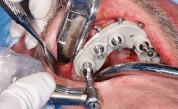
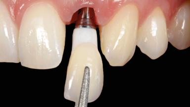
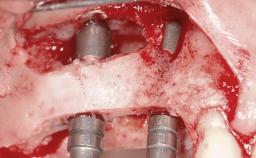
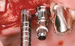
Immediate Implant Placement and Immediate Provisionalization with a Prefabricated-Shell Provisional Crown
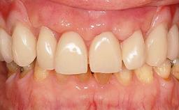
In this case, Arndt Happe describes how he achieved a stable outcome at 5 years by giving careful attention to the coronal aspect of the transmucosal area of the provisional, creating a slim emergence profile.
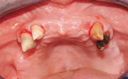
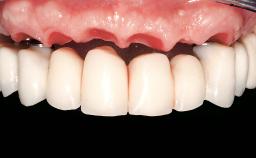
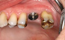
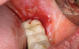
A healthy 31-year-old female patient presented with a failing maxillary left lateral incisor crown. The crown regularly loosened, and the remaining tooth was neither restorable nor rational to treat. The patient had a high smile line, a medium soft tissue biotype with a compromised mesial papilla (shorter than the contralateral one), and a horizontal scar in the buccal soft tissue as a result of past periapical surgery.
Surgical classification
Complex
Prosthodontic classification
Complex
Learner Level
|
1
|
2
|
3
|
Source
Treatment Guide 14
CME/CPD
0.25 hours
Purchase price
10
You must be logged in to access or purchase this item.
There is a wealth of resources you can access by signing up
as an ITI Member. Learn more about ITI membership benefits.
General Risk Assessment
Patient-related Factors
| Smoking Habit | None |
|---|---|
| Oral hygiene | Good |
| Compliance | Good |
| Patient's Expectations | Realistic |
Patient-medical Factors
| Medical Status | Healthy, uneventful healing |
|---|---|
| Medical Fitness | Healthy, able to undergo planned anesthesia and surgical procedure (ASA I) |
| Medications | No medications that would negatively affect the surgical procedure and outcomes. |
| Radiation Treatment | None |
| Growth Status | Complete |
Site-related Factors
| Periodontal Status | No history of periodontal disease, or any active periodontal disease. |
|---|---|
| Access | Adequate |
| Pathology near the implant site | None |
| Previous surgeries in planned implant site | Previous procedures resulting in none or minimal bone and soft tissue changes. |
Surgical Classification
Surgical Complexity
| Timing of placement | Immediate Placement (extraction sockets) (Type I) |
|---|---|
| Simultaneous or Staged grafting procedures | Implant placement with simultaneous hard and soft tissue procedures |
Anatomy
| Bone Volume - Horizontal | Adequate |
|---|---|
| Bone Volume - Vertical | Adequate |
| Keratinized Tissue | Minimal (2-4 mm) |
| Soft Tissue Quality | Presence of minimal scars/no inflammation |
| Proximity to vital anatomic structures | Minimal risk of involvement |
Adjacent Teeth
| Papilla | Deficient |
|---|---|
| Recession | Absent |
| Interproximal attachment | At CEJ |
Extractions
| Radicular morphology | Uniradicular |
|---|---|
| Available apical bone to achieve primary stability | Sufficient height ( ≥ 4 mm) and width (> 2 mm around apex of planned implant) |
| Socket walls | Intact |
| Thickness of buccal wall | less than 2 mm |
| Anticipated residual defect after implant placement | 2 mm or less |
Prosthodontic Classification
Complicating Factors
| Biological | Screw-retained restorations with appropriate contours |
|---|---|
| Mechanical/Technical | Absence of contributing factors |
Prosthesis Factors
| Prosthetic volume | Adequate. Space available for ideal anatomy of the restoration |
|---|---|
| Inter-occlusal space | Adequate. Capable to create an anatomically & functionally correct planned restoration |
| Volume and characteristics of the edentulous ridge (fixed) | Adequate. No adjunctive therapy or prosthetic soft tissue replacement will be necessary |
Esthetic Factors
| Gingival display at full smile | High |
|---|---|
| Shape of tooth crowns | Triangular |
| Restorative status of neighboring teeth | Virgin |
| Gingival Phenotype | Medium-scalloped, medium-thick |
| Bone level on adjacent teeth | ≤5 mm to contact point |
Occlusal Factors
| Occlusal scheme | User-defined occlusal scheme achievable |
|---|---|
| Involvement in occlusion | Minimal or no involvement |
| Occlusal parafunction | Absent |
Complexity
| Loading Protocol | Immediate |
|---|---|
| Implant-supported provisional restoration | Required, elevated esthetic and/or functional demands |
| Timing of placement | Immediate Placement (extraction sockets) (Type I) |
Esthetic Risk Assessment
Esthetic Risk Assessment
| Medical Status | Healthy, uneventful healing |
|---|---|
| Smoking Habit | None |
| Gingival display at full smile | High |
| Width of edentulous span | 1 tooth (≥ 7mm, standard diameter implant) 1 Tooth (≥ 6mm, narrow diameter implant) |
| Shape of tooth crowns | Triangular |
| Restorative status of neighboring teeth | Virgin |
| Gingival Phenotype | Medium-scalloped, medium-thick |
| Infection at implant site | None |
| Soft tissue anatomy | Soft-tissue defects |
| Bone level on adjacent teeth | ≤5 mm to contact point |
| Thickness of buccal wall | less than 2 mm |
| Patient's Expectations | Realistic |
Share this page
Download the QR code with a link to this page and use it in your presentations or share it on social media.
Download QR code